

NEWS AND UPDATES
The latest updates regarding our products and our company

How to Choose the Best Skin Analyzer Device?
Choosing a Skin Analyzer Device is not simply a matter of comparing screen size or counting imaging modes. The right system should fit the people who will use it, the questions they need to answer, and the setting where assessments take place. A salon may value quick, clear consultations, while a clinic may need repeatable measurements and detailed records. Small details matter: Can the camera capture the same cheek area consistently? Does the software explain its scores in plain language? Can staff clean the contact surfaces easily between clients?
Look beyond polished demonstrations. Ask how the device was tested, whether its measurements are repeatable, and what training the supplier provides. Results can shift with room lighting, makeup, hydration, or camera position. That is easy to forget. A useful system should help control these variables, not hide them behind a confident-looking report. Check whether it supports your workflow, stores images securely, and offers dependable technical support. These practical points can matter more than an impressive feature list.
A skin analyzer can support observation and conversation, but it cannot explain every concern or replace professional judgment. Be cautious of claims that promise certainty from a single scan. Even experienced users may need to question a result that does not match what they see. The following guide explores the main technologies, evaluation criteria, and questions to ask before buying, so you can choose a device that is useful, understandable, and suited to real-world use.
What a Skin Analyzer Device Measures and How It Works
How to Choose the Best Skin Analyzer Device?
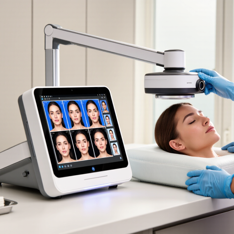
A skin analyzer captures information that a mirror cannot show clearly. Depending on its sensors and imaging modes, it may estimate hydration, surface oil, pore appearance, redness, uneven pigmentation, and skin texture. Some systems use visible light, polarized light, or ultraviolet illumination to highlight different features. The exact measurements vary by device.
Inside, a camera or sensor records images and readings. Software then compares them with reference scales or processes them into visual maps. These results are estimates, not medical diagnoses. Lighting, recent cleansing, makeup, and even room temperature can affect readings. For a fair comparison, analyze clean, dry skin under similar conditions each time. Small changes can be misleading.
Tips: Ask which measurements the device actually takes, and how results are produced. Keep the same distance and lighting during repeat scans. A hydration reading after a hot shower may differ from one taken later. That detail is easy to miss. Treat the report as one source of information, and discuss persistent irritation or sudden skin changes with a qualified healthcare professional. Even good devices have limits, and their scores may not tell the whole story.
How Skin Analyzer Devices Measure the Skin
Many skin analyzers capture images under different lighting conditions and use software to assess visible features such as skin tone, texture, and redness. Some also use ultraviolet light to reveal fluorescence patterns associated with surface oil and pores. Wavelength ranges are approximate; available modes and interpretation vary by device.
How to Define Your Skin Analysis Needs and Intended Use
How to Choose the Best Skin Analyzer Device?
How to Define Your Skin Analysis Needs and Intended Use
Start by naming the questions you need the device to answer. Are you tracking hydration, surface oil, visible redness, pigmentation, or pore appearance? Different systems measure or display different features, so a detailed image does not automatically mean a useful result. Be specific. Decide whether you need a baseline for consultations, repeat measurements over time, or educational images for clients. These uses call for different levels of detail and workflow.
Think about who will operate the device and where. A busy clinic may need quick capture, clear reports, and consistent results across staff. A research setting may require documented methods and more controlled conditions. Consider training, setup time, cleaning, storage, and how results will be explained. For follow-up images, keep lighting, camera distance, and preparation as consistent as possible. Small changes can make comparisons less reliable.
Check what evidence supports the device’s measurements, including its limits across skin tones and conditions. Ask how results are produced and whether the system has been tested for your intended use. A polished image can feel objective. It is not a diagnosis, and it may not answer every clinical question. It is tempting to choose the device with the most features; some may go unused. Write down your essential measurements and daily workflow before comparing specifications.
Which Imaging Technologies and Features to Compare
When comparing skin analyzer devices, look beyond the number of cameras. Standard RGB imaging records visible tone and texture. Cross-polarized light reduces surface glare, making redness and pigment easier to inspect. UV or multispectral imaging may reveal features that ordinary light misses, but results depend on illumination and software. A vivid image is not automatically a reliable one. Not always.
Check whether the device captures consistent images across visits. Ask about fixed camera distance, calibrated lighting, repeatable positioning, and measurement scales. The International Skin Imaging Collaboration’s 2020 challenge dataset included 33,126 dermoscopic images; this research resource shows the scale of image-based analysis, not proof that any analyzer performs equally well. Request validation data for the specific device and the intended skin types.
Useful features include side-by-side progress views, clear reports, exportable images, and secure access controls. Confirm who can review the findings and how uncertain results are flagged. The American Academy of Dermatology estimates that one in five Americans develops skin cancer by age 70. That figure signals the value of careful assessment, not a reason to treat an analyzer as a diagnostic substitute. Test the device under real clinic lighting, with varied skin tones, before relying on its scores. A rushed demo can hide problems.
How to Choose the Best Skin Analyzer Device? - Which Imaging Technologies and Features to Compare
| Technology or Feature | What It Captures or Measures | Common Uses | Advantages | Limitations to Consider | What to Compare Before Buying |
|---|---|---|---|---|---|
| Standardized visible-light imaging | Color photographs of the skin's visible appearance under controlled illumination. | Baseline documentation; visible redness, spots, uneven tone, and general appearance. | Intuitive images that are easy to review and compare over time. | Results can vary with lighting, camera settings, pose, distance, and white balance. A photograph alone does not establish a diagnosis. | Consistent lighting, fixed positioning, repeatable camera settings, and clear image-storage and comparison tools. |
| Cross-polarized imaging | Images captured with crossed polarizing filters that reduce surface glare and can make some color variations beneath the skin surface easier to see. | Documenting visible pigmentation and redness patterns with less reflection from the skin surface. | Reduces specular shine, which can otherwise obscure details in photographs. | Image appearance depends on the light source, filters, and processing; it does not reveal skin structures with diagnostic certainty. | Whether images are repeatable across sessions and whether the system explains how images are processed. |
| Parallel-polarized imaging | Polarized light and camera filters aligned in the same direction, retaining more surface reflection than cross-polarized imaging. | Visualizing surface texture, fine lines, and some skin-surface irregularities. | Can emphasize surface detail and complement other image modes. | Reflections can vary with skin oil, moisture, product residue, and positioning; findings are image-based estimates. | Image consistency, preparation instructions, and whether the feature adds useful information beyond standard photography. |
| UV-induced fluorescence imaging | Fluorescence emitted by certain skin-surface substances when illuminated with ultraviolet or near-ultraviolet light. | Showing some surface features, such as fluorescence associated with porphyrins, and assisting visual skin assessments. | Provides an image mode not visible in ordinary white-light photographs. | Fluorescence can be affected by cosmetics, cleansers, and other surface substances. It is not a standalone test for acne, infection, or other conditions. | Light wavelength, eye-safety guidance, cleaning and preparation protocol, and the intended interpretation of the images. |
| Multispectral imaging | Images acquired at multiple selected wavelength bands, which can provide different information about light absorption and reflection in skin. | Estimating and mapping features associated with pigmentation, redness, or other optical characteristics. | Offers more wavelength-specific information than a single visible-light image. | Outputs depend on wavelength selection, calibration, algorithms, and skin characteristics. A displayed map is an estimate, not a direct measurement of every skin layer. | Available wavelength bands, calibration procedures, validation information, and clear definitions of each reported score or map. |
| 3D imaging or stereophotogrammetry | A three-dimensional surface model reconstructed from multiple images or views. | Assessing facial surface contours, volume changes, and some topographic features over time. | Adds surface-shape information that a flat photograph cannot provide. | Accuracy depends on capture conditions, registration, facial expression, and movement. It does not show internal tissue directly. | Capture time, repeatability, movement tolerance, measurement units, and tools for aligning follow-up scans. |
| Automated image analysis | Software-generated estimates or scores for image features such as visible spots, pores, wrinkles, or redness. | Organizing observations, supporting consultations, and tracking changes in standardized images. | Can make image review more structured and help compare results across visits. | Scores depend on the algorithm, image quality, and population used for validation; different systems may use non-equivalent scales. | Definitions for each score, validation details, export options, and whether results can be reviewed alongside the original images. |
| Calibration and repeatability | Procedures and design features that help keep image capture and measurements consistent between sessions. | Longitudinal tracking and fair comparison of images or scores over time. | Improves the usefulness of follow-up comparisons when capture conditions are controlled. | Consistency does not by itself prove clinical accuracy or make results comparable across different devices. | Routine calibration needs, fixed positioning aids, standardized protocols, and documented repeatability data. |
| Data management and privacy | How images, measurements, and client or patient records are stored, accessed, exported, and protected. | Maintaining records, reviewing progress, and sharing results with authorized users. | Supports organized follow-up and more efficient image comparison. | Cloud storage, access controls, retention, and data-transfer practices differ; privacy obligations depend on location and use. | Consent workflow, encryption, user permissions, backup and deletion policies, export format, and applicable privacy requirements. |
Selection tip: Match the imaging modes to your intended use, prioritize consistent capture and transparent measurement methods, and treat device-generated scores as supportive estimates rather than medical diagnoses.
How to Assess Accuracy, Usability, and Data Privacy
Accuracy begins with the test set, not the screen’s polished score. A 2017 Nature study assessed a deep-learning system using 129,450 clinical images across 2,032 skin conditions. Its results concerned specific image-classification tasks, not every skin analyzer or everyday lighting condition. Ask whether a device was tested on varied skin tones, lighting, and real users. Check sensitivity and specificity, and ask how often results were independently validated. A high score alone is not proof.
Usability shows up in small moments. Can you capture a clear cheek image without repeated prompts? Does the device explain weak lighting, retake limits, and what its readings can and cannot indicate? The 2024 Data Breach Investigations Report found that 68% of breaches involved a non-malicious human element. That matters. Look for simple consent controls, limited data collection, clear deletion options, and safeguards for stored images. Read the privacy policy before uploading a face photo.
Compare the same area under similar lighting, then see whether the result stays consistent. Ask who can access images, how long they are kept, and whether they are shared for secondary uses. A dashboard may feel reassuring. Still, an unexplained score can mislead. I would want plain-language limitations beside each result, not buried in fine print. No device replaces a qualified clinical assessment when a spot changes, bleeds, or persists.
How to Compare Cost, Maintenance, and Manufacturer Support
Compare the purchase price with the full cost of ownership, not the showroom quote. Add software fees, calibration, replacement parts, staff training, shipping, and downtime. Grand View Research valued the global medical aesthetics market at USD 17.1 billion in 2023 and projected 11.5% annual growth through 2030 in its Medical Aesthetics Market report. This growth makes service capacity worth checking, but it does not guarantee reliable support. I still find recurring fees easy to overlook.
Ask how often the device needs calibration and whether maintenance can happen on site. Request written response times, loaner availability, remote diagnostics, and local technician coverage. Check what voids the warranty. A low price can become costly if a camera module fails midweek and no replacement is available. Compare service contracts line by line; vague terms deserve a follow-up question.
Tips: Build a three-year cost sheet and ask the supplier to price common repairs. Request a sample analysis report and a live support demonstration before buying. Note how long image processing takes, and whether staff can repeat the same scan reliably. Keep a little room for surprises.
Article Source:
Schedule
Monday to Thursday:
8:15 – 17:00
Friday:
8:00-15:00
Address
Leonardo Da Vinci, 10
Valencia Technology Park
46980 Paterna, Valencia, SPAIN
Real Laboratory SL
info@site_e1c8a3bd-d539-4d7f-8695-37f21373de39
Teléfono: +34 96 136 61 07
